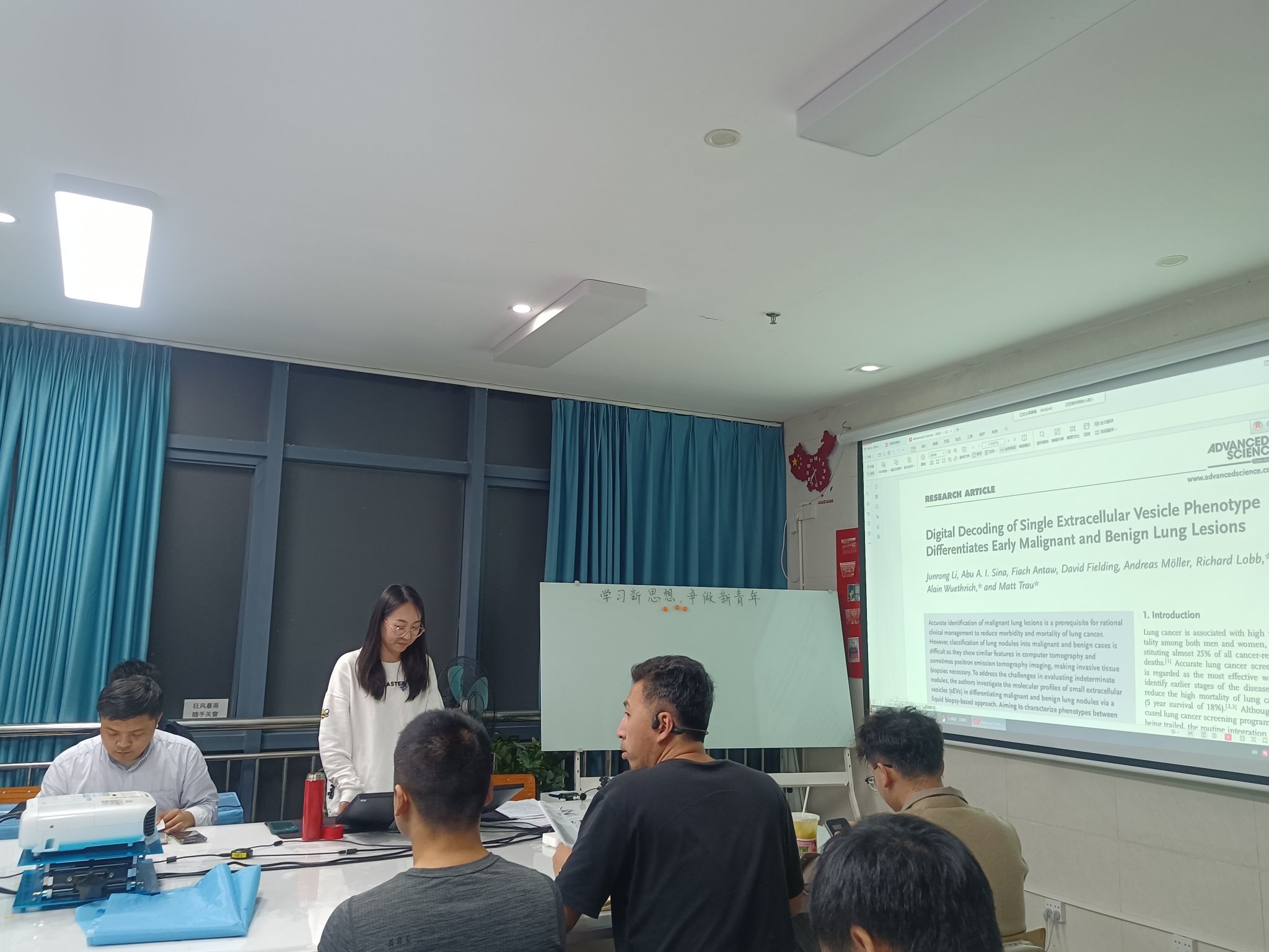
Digital Decoding of Single Extracellular Vesicle Phenotype Differentiates Early Malignant and Benign Lung Lesions
单个细胞外囊泡表型的数字解码区分早期恶性和良性肺部病变

主讲人:唐静施
https://doi.org/10.1002/advs.202204207
Adv Sci (Weinh) 2022 11 17
Abstract:
differentiating malignant and benign lung nodules via a liquid biopsy-based approach. Aiming to characterize phenotypes between malignant and benign groups, they develop a single-molecule-resolution-digital-sEV-counting-detection (DECODE) chip that interrogates three lung-cancer-associated sEV biomarkers and a generic sEV biomarker to create sEV molecular profiles. DECODE capturess EVs on a nanostructured pillar chip, confines individual sEVs, and profiles sEV biomarker expression through surface-enhanced Raman scattering barcodes.The author utilize DECODE to generate a digitally acquired sEV molecular profiles in a cohort of 33 people, including patients with malignant and benign lung nodules, and healthy individuals.Significantly, DECODE reveals sEV-specific molecular profiles that allow the separation of malignant from benign (area under the curve, AUC = 0.85), which is promising for non-invasive characterisation of lung nodules found in lung cancer screening and warrants further clinincal validaiton with larger cohorts.
摘要:
准确识别肺部恶性病变是合理临床治疗的前提,以降低肺癌的发病率和死亡率。然而,将肺结节分类为恶性和良性病例是困难的,因为在计算机断层扫描和正电子发射计算机断层扫描成像中显示相似的特征,因此进行侵入性组织活检是必要的。为了解决评估不确定结节的挑战,作者通过基于液体活检的方法研究了细胞外小泡(sev)在区别恶性和良性肺结节中的分子图谱。为了解决评估不确定结节的挑战,作者通过基于液体活检的方法,研究了小细胞外囊泡(sev)在区分恶性和良性肺结节中的分子谱。为了表征恶性和良性组之间的表型,他们开发了一种单分子分辨率数字小细胞外囊泡计数检测芯片(DECODE),可以研究三种肺癌相关的小细胞外囊泡生物标志物和一种通用的小细胞外囊泡标志物,以创建小细胞外囊泡分子谱。DECODE在纳米结构柱芯片上捕获小细胞外囊泡,限制单个小细胞外囊泡,并通过表面增强拉曼散射条形码来表达小细胞外囊泡标志物图谱。作者利用DECODE生成一个数字获取小细胞外囊泡分子谱,在一个包括恶性和良性肺结节患者以及健康个体的33人的队列中。值得注意的是,DECODE揭示了细胞外囊泡特异性分子谱,可以将恶性和良性进行区分(曲线下面积,AUC=0.85),这有望用于在肺癌筛查中发现肺结节非侵入性特征,并需要在更大的队列中进一步进行临床验证。

